The thoracic cage – the rib cage and sternum
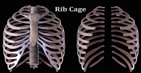
Data Rib cage
There is one final part of the hub skeleton we didn’t cover last lab: the thoracic enclosure, likewise called the rib confine. The thoracic enclosure encompasses and secures the heart and lungs in the thoracic depression. It comprises of the ribs, the sternum, and the thoracic vertebrae, to which the ribs articulate.
We inspected the thoracic vertebrae last lab, so here we will just analyze the ribs and sternum.
There are twelve sets of ribs. The number is something similar in the two guys and females. Each pair expresses with an alternate thoracic vertebra on the back side of the body. The most unrivaled rib is assigned rib 1 and it verbalizes with the T1 thoracic vertebrae. The rib underneath that is rib 2, and it interfaces with the T2 thoracic vertebra, etc. Ten of the twelve ribs interface with pieces of hyaline ligament on the front side of the body. The ligament strips are called costal ligament (“costal” is the physical modifier that alludes to the rib) and associate on their opposite finish to the sternum.
On a singular rib, one end has different cycles, features, and knocks. This is the end that expresses with the vertebra. The opposite end is gruff and smooth. This is the end that interfaces with costal ligament (except if it is a drifting rib. See underneath.)
Ribs 1-7 are known as the genuine ribs. Each obvious rib cage interfaces with its own portion of costal ligament, which thusly associates with the sternum. Ribs 8-12 are known as the bogus ribs. Ribs 8, 9, and 10 do interface with costal ligament, yet the costal ligament of every one of these ribs associates with the costal ligament of the rib above it, rather than straightforwardly to the sternum. Ribs 11 and 12 don’t have any costal ligament associated with them whatsoever, and as well as being gathered in the bogus ribs, these two are additionally called drifting ribs, to mirror that reality.
The sternum has three sections. The manubrium, at the prevalent finish of the sternum, and more extensive than the remainder of the bone, gives explanation focuses to the clavicles and for the costal ligament stretching out from rib 1. The focal, slender body gives explanation focuses to costal ligament from rib cage 2 through 7. The xiphoid cycle which hangs down at the second rate end of the interaction (“xiphoid” is from the Greek for blade), begins as ligament, and doesn’t commonly solidify into bone until an individual is around 40 years of age.
The ribcage upholds the chest area, secures inward organs, including the heart and lungs, and helps with relaxing. Rib wounds incorporate injuries, torn ligament and bone cracks. Chest injury may likewise cause dangerous wounds like a penetrated lung or a cracked aorta.
Normal reasons for rib injury incorporate engine vehicle mishaps and falls. Treatment intends to assuage torment while the injury recuperates.
Structure of the ribs
The ribcage consists of 24 curved ribs arranged in 12 pairs. Each pair is attached to a vertebra in the spine. At the front of the body, the first seven pairs of ribs are attached directly to the sternum (breastbone) by cartilage known as costal cartilage. These ribs are often called ‘true ribs’.
The next three pairs of ribs aren’t connected to the sternum. Instead, costal cartilage attaches these ‘false ribs’ to the last pair of true ribs. The remaining two pairs aren’t attached at the front of the body at all and are known as ‘floating ribs’.
The ribcage is supported by ligaments and muscles, including the muscles between the ribs (intercostal muscles). These muscles allow the ribcage to expand when you breathe in and to drop when you breathe out.