The Ribs anatomy of the bones
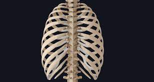
The rib cage are a bunch of twelve combined bones which structure the defensive ‘confine’ of the chest. They articulate with the vertebral section posteriorly, and end anteriorly as ligament (known as costal ligament).
As a feature of the hard chest, the ribs secure the inward thoracic organs. They likewise play a part in ventilation; moving during chest extension to empower lung expansion.
In this article, we will check out the life structures of the ribs – their hard milestones, explanations and clinical connections.
Rib Structure
There are two arrangements of ribs – abnormal and run of the mill. The normal ribs have a summed up structure, while the abnormal ribs have minor departure from this design.
Common Ribs
The common rib comprises of a head, neck and body:
The head is wedge molded, and has two articular features isolated by a wedge of bone. One aspect expresses with the mathematically relating vertebra, and the other verbalizes with the vertebra above.
The neck contains no hard prominences, yet basically associates the head with rib cage the body. Where the neck meets the body there is a roughed tubercle, with a feature for enunciation with the cross over course of the relating vertebra.
The body, or shaft of the rib is level and bended. The interior surface of the shaft has a section for the neurovascular supply of the chest, shielding the vessels and nerves from harm.
Abnormal Ribs
Ribs 1, 2, 10 11 and 12 can be portrayed as ‘abnormal’ – they have highlights that are not normal to every one of the ribs.
Rib 1 is more limited and more extensive than different ribs. It just has one aspect rib cage on its head for explanation with its relating vertebra (there is certifiably not a thoracic vertebra above it). The predominant surface is set apart by two furrows, which clear a path for the subclavian vessels.
Rib 2 is more slender and longer than rib 1, and has two articular features on the head as typical. It has a roughened region on its upper surface, from which the serratus front muscle begins.
Rib 10 just has one aspect – for explanation with its mathematically relating vertebra.
Ribs 11 and 12 have no neck, and just hold back one aspect, which is for explanation with their relating vertebra.
Back
Every one of the twelve ribs articulate posteriorly with the vertebra rib cage of the spine. Each rib structures two joints:
Costotransverse joint – Between the tubercle of the rib, and the cross over costal feature of the relating vertebra.
Costovertebral joint – Between the top of the rib, predominant costal aspect of the relating vertebra, and the mediocre costal feature of the vertebra above.
Front
The front connection of the ribs shift:
Ribs 1-7 append autonomously to the sternum.
Ribs 8 – 10 append to the costal ligaments better than them.
Ribs 11 and 12 don’t have a front connection and end in the stomach muscle structure. Along these lines, they are now and again called ‘drifting ribs’.
Rib breaks most normally happen in the center ribs, as a result of devastating wounds or direct injury. A typical entanglement of a rib crack is further delicate tissue injury from the messed up sections. Designs most in danger of harm are the lungs, spleen or stomach.
On the off chance that at least two cracks happen in at least two neighboring ribs, the impacted region is presently not taken care of the thoracic muscles. It shows a confusing development during lung swelling and flattening. This condition is known as thrash chest. It hinders full extension of the ribcage, subsequently influencing the oxygen content of the blood. Thrash chest is treated by fixing the impacted ribs, forestalling their incomprehensible development.